
Nöroşirürjide cerrahi mikroskopların uygulama geçmişi ve rolü
Nöroşirürji tarihinde, uygulama alanıcerrahi mikroskoplarBu, geleneksel nöroşirürji döneminde çıplak gözle yapılan ameliyatlardan, modern nöroşirürji döneminde ultrason eşliğinde yapılan ameliyatlara geçişi simgeleyen çığır açıcı bir semboldür.mikroskopKim ve ne zaman yaptı?ameliyat mikroskoplarıNöroşirürjide kullanılmaya başlanmasının sebebi nedir? Ne gibi bir rolü var?cerrahi mikroskopNöroşirürjinin gelişiminde hangi rol oynadı? Bilim ve teknolojinin ilerlemesiyle birlikte,Ameliyat mikroskobuDaha gelişmiş ekipmanlarla mı değiştirilmeli? Bu, her beyin cerrahının farkında olması ve en son teknolojiyi ve aletleri beyin cerrahisi alanına uygulayarak beyin cerrahisi becerilerinin geliştirilmesini teşvik etmesi gereken bir sorudur.
1. Tıp Alanında Mikroskopi Uygulamalarının Tarihçesi
Fizikte, gözlük camları, büyütme etkisi olan ve büyütme oranı sınırlı olan, tek bir yapıya sahip dışbükey camlardır ve büyüteç olarak bilinirler. 1590 yılında, iki Hollandalı, ince bir silindirik gövdenin içine iki dışbükey cam plaka yerleştirerek dünyanın ilk kompozit yapılı büyütme cihazını icat ettiler:mikroskopDaha sonra mikroskobun yapısı sürekli olarak geliştirildi ve büyütme oranı sürekli olarak arttı. O zamanlar bilim insanları ağırlıklı olarak bunu kullanıyordu.kompozit mikroskopHayvan ve bitkilerin hücre yapısı gibi minik yapılarını gözlemlemek için. 19. yüzyılın ortalarından sonlarına doğru, büyüteçler ve mikroskoplar tıp alanında giderek daha fazla kullanılmaya başlandı. Başlangıçta cerrahlar, ameliyat için burun köprüsüne yerleştirilebilen tek mercekli yapıya sahip gözlük tipi büyüteçler kullanıyorlardı. 1876'da Alman doktor Saemisch, bileşik gözlük tipi büyüteç kullanarak dünyanın ilk "mikroskobik" ameliyatını gerçekleştirdi (ameliyatın türü bilinmiyor). 1893'te Alman şirketi Zeiss,binoküler mikroskopEsas olarak tıp laboratuvarlarında deneysel gözlem için ve oftalmoloji alanında kornea ve ön kamara lezyonlarının gözlemlenmesi için kullanılır. 1921'de, hayvanların iç kulak anatomisi üzerine yapılan laboratuvar araştırmalarına dayanarak, İsveçli kulak burun boğaz uzmanı Nylen sabit bir cihaz kullandı.tek gözlü cerrahi mikroskopNylen'in kronik orta kulak iltihabı ameliyatlarını insanlarda gerçekleştirmek için tasarladığı ve ürettiği, gerçek bir mikrocerrahi olan bir cihaz. Bir yıl sonra, Nylen'in üstü olan doktor Hlolmgren bir cihazı tanıttı.binoküler cerrahi mikroskopZeiss tarafından ameliyat odasında üretilmiştir.
ErkenAmeliyat mikroskoplarıZayıf mekanik stabilite, hareket ettirilememesi, farklı eksenlerin aydınlatılması ve objektif lensin ısınması, dar cerrahi büyütme alanı gibi birçok dezavantajı vardı. Bunların hepsi, daha geniş uygulama alanını sınırlayan nedenlerdir.cerrahi mikroskoplarSonraki otuz yıl içinde, cerrahlar ve diğer uzmanlar arasındaki olumlu etkileşim sayesindemikroskop üreticileri, performansıcerrahi mikroskoplarsürekli olarak geliştirildi vebinoküler cerrahi mikroskoplar, çatıya monte mikroskoplarZoom lensler, koaksiyel ışık kaynağı aydınlatması, elektronik veya su basıncıyla kontrol edilen mafsallı kollar, ayak pedalı kontrolü ve benzeri özellikler sırasıyla geliştirildi. 1953 yılında Alman şirketi Zeiss, bir dizi özel ürün üretti.kulak hastalıkları için cerrahi mikroskoplarÖzellikle orta kulak ve temporal kemik gibi derin lezyonlara yönelik ameliyatlar için uygundur. Performansı ise...cerrahi mikroskoplarGelişmeye devam ettikçe, cerrahların zihniyeti de sürekli değişiyor. Örneğin, Alman doktorlar Zollner ve Wullstein şunu şart koşmuşlardır:cerrahi mikroskoplarTimpanik zar şekillendirme ameliyatı için kullanılmalıdır. 1950'lerden beri, göz doktorları göz muayenelerinde yalnızca mikroskop kullanma uygulamasını kademeli olarak değiştirmiş ve çeşitli yöntemleri kullanmaya başlamışlardır.otocerrahi mikroskoplarıGöz cerrahisine yöneldi. O zamandan beri,Ameliyat mikroskobuKulak burun boğaz ve göz hastalıkları alanlarında yaygın olarak kullanılmaktadır.
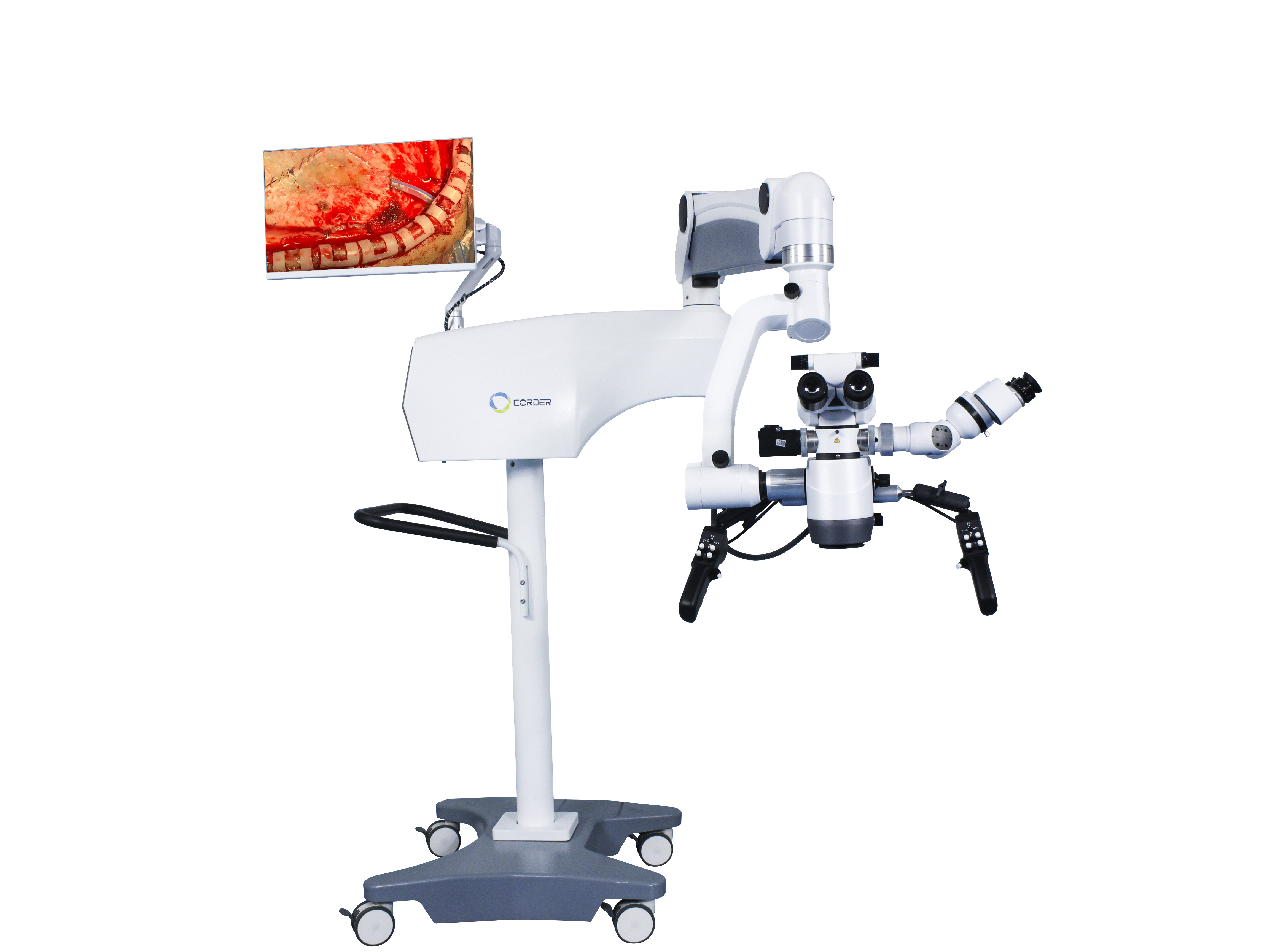
2. Nöroşirürjide cerrahi mikroskobun uygulaması
Nöroşirürjinin kendine özgü yapısı nedeniyle, uygulamasınöroşirürjide cerrahi mikroskoplarKulak burun boğaz ve göz hastalıklarına göre biraz daha geç bir dönemde ortaya çıkmış olup, beyin cerrahları bu yeni teknolojiyi aktif olarak öğrenmektedirler. O dönemde,cerrahi mikroskopların kullanımıEsas olarak Avrupa'daydı. Amerikalı göz doktoru Perrit ilk kez tanıttı.cerrahi mikroskoplar1946'da Avrupa'dan Amerika Birleşik Devletleri'ne gelen bu girişim, Amerikalı nörocerrahların kullanabileceği yöntemlerin temelini attı.Ameliyat mikroskopları.
İnsan hayatının değerine saygı açısından bakıldığında, insan vücudu için kullanılan her yeni teknoloji, ekipman veya alet, öncelikle hayvanlar üzerinde deneylere tabi tutulmalı ve operatörler için teknik eğitim verilmelidir. 1955'te Amerikalı nörocerrah Malis, hayvanlar üzerinde beyin ameliyatı yaparken bir yöntem kullandı.binoküler cerrahi mikroskopAmerika Birleşik Devletleri'ndeki Güney Kaliforniya Üniversitesi'nde beyin cerrahı olan Kurze, kulak ameliyatlarını mikroskop altında gözlemledikten sonra bir yıl boyunca laboratuvarda mikroskop kullanma cerrahi tekniklerini öğrendi. Ağustos 1957'de, 5 yaşında bir çocuğa akustik nöroma ameliyatını başarıyla gerçekleştirdi.kulak ameliyatı mikroskobuBu, dünyanın ilk mikrocerrahi ameliyatıydı. Kısa bir süre sonra Kurze, bir çocuk üzerinde yüz siniri ile dil altı siniri arasında başarılı bir anastomoz gerçekleştirdi; bu işlemi bir mikrocerrahi aleti kullanarak yaptı.cerrahi mikroskopVe çocuğun iyileşmesi mükemmeldi. Bu, dünyadaki ikinci mikrocerrahi ameliyattı. Sonrasında Kurze, çocuğu taşımak için kamyonlar kullandı.Ameliyat mikroskoplarıMikrocerrahi nörocerrahi için çeşitli yerlere başvurdu ve kullanımını şiddetle tavsiye etti.cerrahi mikroskoplarDiğer beyin cerrahlarına. Ardından Kurze, bir yöntem kullanarak beyin anevrizması klipsleme ameliyatı gerçekleştirdi.cerrahi mikroskop(Ne yazık ki, hiçbir makale yayınlamadı.) Tedavi ettiği bir trigeminal nevralji hastasının desteğiyle, 1961'de dünyanın ilk mikro kafa tabanı nöroşirürji laboratuvarını kurdu. Kurze'nin mikrocerrahiye katkısını her zaman hatırlamalı ve yeni teknolojileri ve fikirleri kabul etme cesaretinden ders çıkarmalıyız. Ancak, 1990'ların başlarına kadar Çin'deki bazı nöroşirürjistler bunu kabul etmedi.Nöroşirürji mikroskoplarıAmeliyat için. Bu, bizim için bir sorun değildi.Nöroşirürji mikroskobuSorun bizzat kendisinde değil, beyin cerrahlarının ideolojik anlayışında yatıyor.
1958'de Amerikalı nörocerrah Donaghy, Burlington, Vermont'ta dünyanın ilk mikrocerrahi araştırma ve eğitim laboratuvarını kurdu. İlk aşamalarda, üstlerinden kaynaklanan kafa karışıklığı ve mali zorluklarla da karşılaştı. Akademik hayatta, her zaman beyin trombozu olan hastalardan doğrudan trombüsleri çıkarmak için kortikal kan damarlarını kesmeyi hayal ediyordu. Bu nedenle, hayvan ve klinik araştırmalar konusunda vasküler cerrah Jacobson ile işbirliği yaptı. O zamanlar, çıplak gözle bakıldığında, yalnızca çapı 7-8 milimetre veya daha fazla olan küçük kan damarları dikilebiliyordu. Daha ince kan damarlarının uçtan uca anastomozunu sağlamak için Jacobson önce gözlük tipi bir büyüteç kullanmayı denedi. Kısa süre sonra, birkulak burun boğaz cerrahi mikroskobuJacobson, asistan doktor olduğu dönemde cerrahi alanında kullanmıştı. Bu nedenle, Almanya'daki Zeiss firmasının yardımıyla çift operatörlü bir cerrahi mikroskop tasarladı.Diploskop(Vasküler anastomoz için, iki cerrahın ameliyatı eş zamanlı olarak gerçekleştirmesine olanak tanıyan) bir yöntem geliştirdi. Kapsamlı hayvan deneylerinden sonra Jacobson, köpeklerde ve karotis dışı arterlerde mikrocerrahi anastomoz üzerine bir makale yayınladı (1960), bu makalede vasküler anastomozun %100 açıklık oranı elde edildi. Bu, mikrocerrahi nörocerrahi ve vasküler cerrahi ile ilgili çığır açan bir tıp makalesidir. Jacobson ayrıca mikro makaslar, mikro iğne tutucular ve mikro alet sapları gibi birçok mikrocerrahi alet tasarladı. 1960 yılında Donaghy, bir serebral arter insizyonu trombektomisini başarıyla gerçekleştirdi.cerrahi mikroskopBeyin trombozu olan bir hasta için. Amerika Birleşik Devletleri'nden Rhoton, 1967'de mikroskop altında beyin anatomisini incelemeye başladı, mikrocerrahi anatomi alanında öncülük etti ve mikrocerrahinin gelişimine önemli katkılarda bulundu. Avantajları nedeniylecerrahi mikroskoplarMikrocerrahi aletlerin gelişmesiyle birlikte, giderek daha fazla cerrah bu aletleri kullanmayı tercih etmektedir.cerrahi mikroskoplarCerrahi alanında uzmanlaşmıştır. Ayrıca mikrocerrahi prosedürler üzerine birçok makale yayınlamıştır.
3. Çin'de nöroşirürjide cerrahi mikroskobun uygulaması
Japonya'da yaşayan vatansever bir Çinli olan Profesör Du Ziwei, ilk yerli bağışı gerçekleştirdi.nöroşirürjik mikroskopve ilgilimikrocerrahi aletler1972'de Suzhou Tıp Fakültesi Bağlı Hastanesi Nöroşirürji Bölümü'ne (şimdiki Suzhou Üniversitesi Bağlı Birinci Hastanesi Nöroşirürji Bölümü) katıldı. Çin'e döndükten sonra ilk olarak intrakraniyal anevrizmalar ve menenjiomlar gibi mikrocerrahi ameliyatlar gerçekleştirdi. Mevcut olanaklar hakkında bilgi edindikten sonra...nöroşirürjik mikroskoplarMikrocerrahi aletlerin kullanımını gözlemlemek amacıyla, Pekin Yiwu Hastanesi Nöroşirürji Bölümü'nden Profesör Zhao Yadu, Suzhou Tıp Fakültesi'nden Profesör Du Ziwei'yi ziyaret etti.cerrahi mikroskoplarŞanghay Huashan Hastanesi'nden Profesör Shi Yuquan, mikrocerrahi işlemlerini gözlemlemek üzere Profesör Du Ziwei'nin bölümünü bizzat ziyaret etti. Bunun sonucunda, mikrocerrahi yöntemlerinin tanıtılması, öğrenilmesi ve uygulanmasında bir dalga başladı.Nöroşirürji mikroskoplarıÇin'deki büyük nöroşirürji merkezlerinde ortaya çıkan bu gelişme, Çin'in mikro nöroşirürjisinin başlangıcını işaret etti.
4. Mikrocerrahi Ameliyatının Etkisi
Kullanımından dolayınöroşirürjik mikroskoplarÇıplak gözle yapılamayan ameliyatlar, 6-10 kat büyütme koşullarında mümkün hale gelir. Örneğin, etmoidal sinüs yoluyla hipofiz tümörü ameliyatı yapmak, normal hipofiz bezini korurken hipofiz tümörlerini güvenli bir şekilde tespit edip çıkarmayı sağlar; çıplak gözle yapılamayan ameliyatlar, beyin sapı tümörleri ve omurilik içi tümörleri gibi daha iyi ameliyatlar haline gelir. Akademisyen Wang Zhongcheng, büyütme kullanılmadan önce serebral anevrizma ameliyatında %10,7'lik bir ölüm oranına sahipti.nöroşirürji mikroskobu1978'de mikroskop kullanıldıktan sonra ölüm oranı %3,2'ye düştü. Mikroskop kullanılmadan yapılan serebral arteriyovenöz malformasyon ameliyatlarının ölüm oranı...cerrahi mikroskopBu oran %6,2 idi ve 1984'ten sonra, bir yöntemin kullanımıyla...nöroşirürji mikroskoplarıÖlüm oranı %1,6'ya düştü. Kullanımıyla birliktenöroşirürji mikroskobuBu yöntem, kraniotomiye gerek kalmadan, minimal invaziv transnazal transsfenoidal yaklaşım yoluyla hipofiz tümörlerinin tedavi edilmesini sağlayarak cerrahi mortalite oranını %4,7'den %0,9'a düşürmektedir. Bu sonuçlara geleneksel kaba göz cerrahisi ile ulaşılamaz, bu nedenlecerrahi mikroskoplarModern nöroşirürjinin sembolüdürler ve modern nöroşirürjide vazgeçilmez ve yeri doldurulamaz cerrahi ekipmanlardan biri haline gelmişlerdir.
Yayın tarihi: 09-12-2024


